BIOS 255 Week 2 Concepts I The Cardiovascular System- Heart
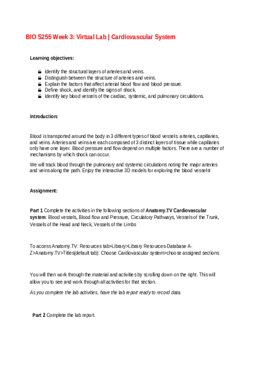
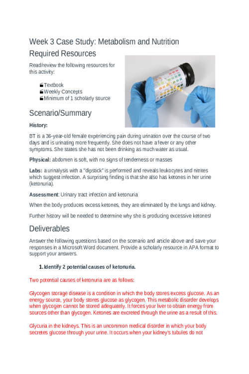
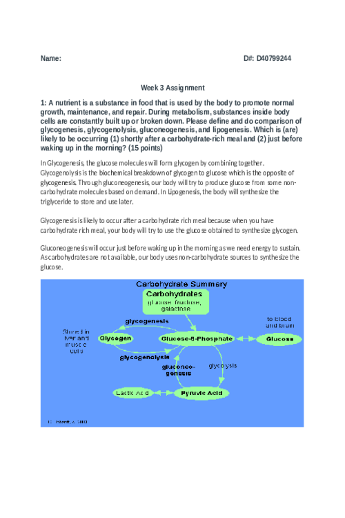
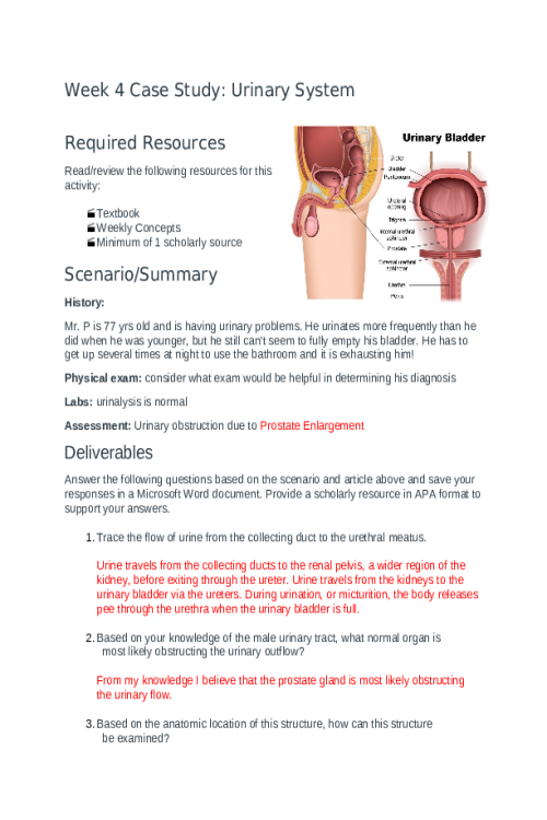
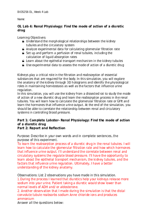
Gross and Microscopic Anatomy of the Heart Introduction The heart represents the central organ that we associate with the cardiovascular system. This organ is associated with two distinct sets of blood vessels—pulmonary and systemic. Before delving into The Position of the Heart in the Thoracic Cavity these circulatory systems, you must first learn about the gross anatomical features of this four-chambered hydraulic pump. In the first week of the course, we learned about the properties of blood including its development, function, and appearances. We are going to shift our focus this week to the mechanisms of pumping the blood conducted by the heart. The heart is in the mediastinum of the thoracic cavity surrounded by loose connective tissue known as the pericardium. The majority of the heart rests to the left side of the sternum, pointing into the left lung between ribs 2 and 5. If you feel (palpate) the left side of the chest, you should be able to feel rhythmic contractions. If we were to observe the heart, we would see that it comes to a rounded point. This structure is known as the apex and it rests just above the diaphragm. Pericardium As mentioned, the heart is surrounded by a connective tissue serous membrane known as the pericardium. Peri refers to "around" and cardio refers to "heart." Outside the serous pericardium a thicker connective tissue layer can be found that is called the fibrous pericardium. Previously you learned that serous membranes would have two sides. The side that faces the heart is known as the visceral pericardium. Visceral refers to organs or organ-side. The side that faces away from the heart is known as the parietal pericardium. Parietal refers to the side of a structure or facing away. We have seen the word parietal when referring to the parietal bone and parietal lobe, both of which are located on the sides of the skull and brain, respectively. Between the parietal and visceral layers of serous membranes is typically a volume of serous fluid. The pericardial fluid that resides within the pericardium acts to reduce friction as the heart beats. Layers of the Heart Wall Moving deeper in the heart, there are 3 distinct layers: the epicardium, the myocardium, and finally, the innermost layer, the endocardium. The epicardium is the outer-most layer that consists of the visceral pericardium and is made of the mesothelium, adipose tissue, and elastic fibers. This layer functions in the production of pericardial fluid and reduction of friction against the surrounding tissues. The myocardium is the thick middle layer that consists of the cardiac muscle. This layer functions in pumping blood throughout the body to deliver nutrients and oxygen. The endocardium is the inner-most layer of the heart. It consists of an epithelial layer and an underlying basement membrane and is similar to the endothelium of blood vessels. This layer functions in providing a smooth surface for blood movement, protection of the valves, and regulating contractility. The Atria and Ventricles Looking at the internal structures of the heart, 4 distinct chambers are visible, 2 superior atria and 2 inferior ventricles. Separating the left and right sides of the heart is thick layer of myocardium known as the septum. The walls of the atria are thin and function to collect blood from the major veins of the body and deliver it to the ventricles in a controlled manner. 9540851335980The walls of the ventricles are significantly thicker with myocardium which allows them to generate enough force (pressure) to pump blood through the pulmonary and systemic circuits. The left ventricular wall is significantly thicker because it must pump blood to the entire body while the right ventricle needs only to deliver blood to the lungs and back. The Surface of the Heart When determining which side is left and which is right, the apex, or the point of the heart, will be pointing towards the left of the atrium. Thus the left atrium will appear superiorly and to the patient’s left (the provider’s right). The ventricles will be found inferior to the atria, close to the apex. 978295848300The superior portion of each atrium is a flap or ’ear-like’ region known as the auricle that acts as a small blood reservoir to receive blood from their respective vessels. The Cardiac Valves The function of the atrioventricular valves is to prevent backflow from the ventricles into the atria. Tricuspid valve: Separates the right atrium from the right ventricle. It consists of three flaps and allows deoxygenated blood to flow from the right atrium to right ventricle. Bicuspid/mitral valve: Separates the left atrium from the left ventricle. It consists of two flaps and allows oxygenated blood to flow from the left atrium to the left ventricle. The function of the semilunar valves is to prevent backflow from the aorta and pulmonary trunk into the ventricles. Pulmonary semilunar valve: Separates the right ventricle from the pulmonary trunk. Aortic semilunar valve: Separates the left ventricle from the aorta. 970094153161The Primary Internal Structures of the Heart Though we have been focusing on the major anatomical structures of the heart, we still need to observe the smaller anatomical structures that will allow for additional functionality. The interventricular septum is an area of myocardium that separates the left and right ventricles of the heart to prevent the mixing of oxygenated and deoxygenated blood. Similarly, there is an interatrial septum that divides the left and right atria. Papillary muscles are found on the internal surface of the ventricles and connect to the atrioventricular valves by tendinous threads called chordae tendineae. During ventricular contraction, papillary muscles also contract and pull on the chordae tendineae to prevent the atrioventricular valves from everting into the atria and allowing blood to flow backwards (known as tricuspid/bicuspid valve regurgitation). 963799870525Blood Flow To and From the Heart Wall The fibrous skeleton consists of a high-density area of connective tissue that provides anchorage for the valves and provides electrical insulation between the atria and the ventricles. For a tissue that is constantly contracting, it will need its own supply of oxygen, and the heart is no exception! The blood will not be able to deliver oxygen to the myocardium via the chambers, rather it will require unique blood vessels to deliver the oxygenated blood. The right and left coronary arteries branch off from the ascending aorta. The right coronary artery branches to form the posterior interventricular branch and the marginal branch whereas the left coronary artery branches to form the anterior interventricular branch. This means the right coronary artery will move towards posterior aspects of the heart and the left coronary artery will move towards the anterior aspects. Once the oxygen has been off loaded, the deoxygenated blood will be drained into the great cardiac vein, middle
Related Products
BIOS 242 Week 1 Assignment; Lab 1 of 14 Onsite - Culture Transfer Techniques
Contributor: Matthew Lillard
$10.00
BIOS 242 Week 1 Assignment; Lab 2 of 14 Onsite - Isolation of Pure Cultures
Contributor: Matthew Lillard
$10.00
BIOS 242 Week 2 Assignment; Lab 3 of 14 Onsite; Microscopy Observation of Cells
Contributor: Matthew Lillard
$10.00
BIOS 242 Week 2 Discussion; Characteristics of Prokaryotes and Eukaryotes Cells
Contributor: Matthew Lillard
$10.00
BIOS 242 Week 2 Assignment; Concepts; Classification and Nutrition
Contributor: Matthew Lillard
$10.00
BIOS 242 Week 3 Assignment; Lab 6 of 14 Onsite; Carbohydrate Fermentation
Contributor: Matthew Lillard
$10.00
BIOS 242 Week 3 Discussion; Culturing Microorganisms - Halobacterium noricense
Contributor: Matthew Lillard
$10.00
BIOS 242 Week 3 EXAM 1. Chapters 1, 3, 4, 5, 6, 9 - Multiple Choice Review Questions
Contributor: Matthew Lillard
$10.00
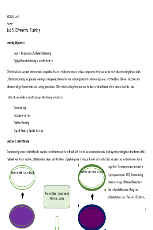
BIOS 242 Week 3 Assignment; Lab 5 of 14 Onsite; Differential Staining
Contributor: Matthew Lillard
$10.00
BIOS 242 Week 5 Assignment; Lab 10 of 14 Onsite; How Germs Spread..
Contributor: Matthew Lillard
$10.00
BIOS 242 Week 5 Assignment; Lab 9 of 14 Onsite; Food Safety.docx
Contributor: Matthew Lillard
$10.00
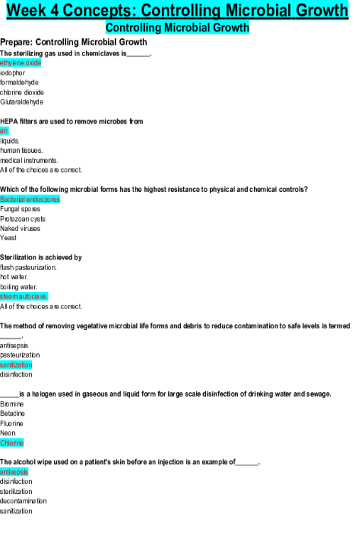
BIOS 242 Week 4 Assignment; Concepts; Controlling Microbial Growth
Contributor: Matthew Lillard
$10.00
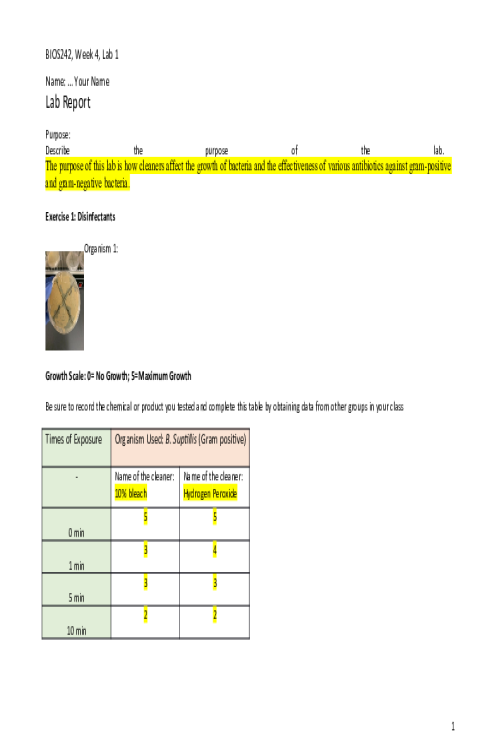
BIOS 242 Week 4 Assignment; Lab 7 of 14 Onsite; Disinfectants and Antibiotics
Contributor: Matthew Lillard
$10.00
BIOS 242 Week 4 Assignment; Lab 8 of 14 Onsite; Fomite Transmission
Contributor: Matthew Lillard
$10.00
BIOS 242 Week 6 Assignment; Pick Your Pathogen - Clostridium Perfringens
Contributor: Matthew Lillard
$10.00
BIOS 242 Week 6 Assignment; Concepts; Epidemiology of Infectious Diseases
Contributor: Matthew Lillard
$10.00
BIOS 242 Week 6 Assignment; Lab 11 0f 14 Onsite; Bacteria of the skin
Contributor: Matthew Lillard
$10.00
BIOS 242 Week 6 Assignment; Lab 12 0f 14 Onsite; Bacteria of Respiratory System
Contributor: Matthew Lillard
$10.00
BIOS 242 Week 7 Assignment; Lab 14 of 14 Onsite; Unknown Bacteria
Contributor: Matthew Lillard
$10.00
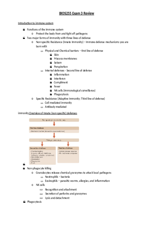
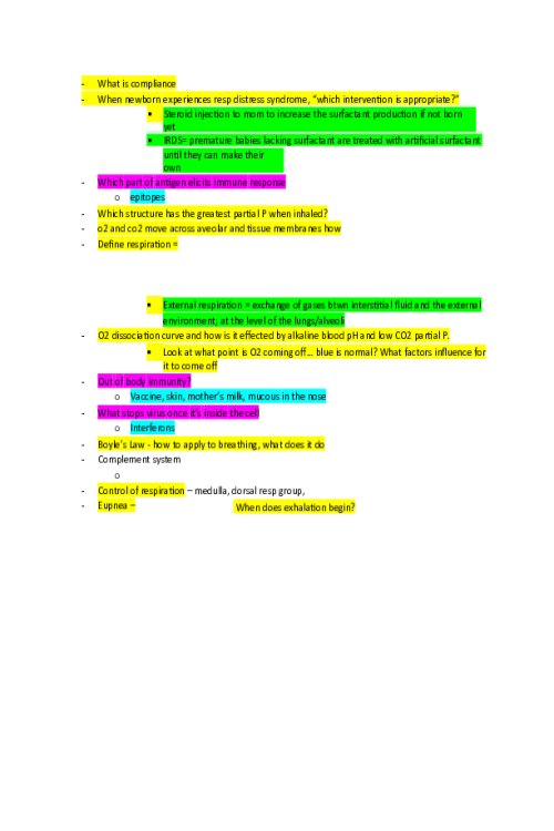
BIOS 242 Week 8 EXAM 3 Review. Chapters 14-23 (Excluding 16, 17)
Contributor: Matthew Lillard
$10.00
BIOS 242 Week 7 Assignment; Lab 13 of 14 Onsite; Bacteria of Digestive System
Contributor: Matthew Lillard
$10.00
BIOS 255 Week 1 Virtual Lab; Hematology Part I, Part II, Part III
Contributor: Chandler Hallow
$10.00
BIOS 255 Week 2 Virtual Lab; Cardiovascular Function during Exercise Part 1, Part II, Part III
Contributor: Chandler Hallow
$10.00
BIOS 255 Week 3 Concepts I The Cardiovascular System- Blood Vessels (Quiz)
Contributor: Matthew Lillard
$10.00
BIOS 255 Week 4 Assignment; Case Study I Natural Killer Cell Deficiency
Contributor: Matthew Lillard
$10.00
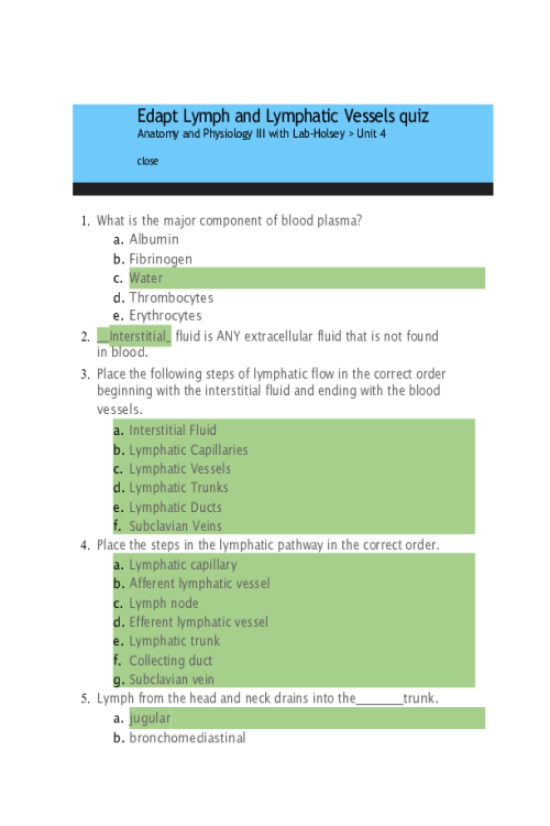
BIOS 255 Week 4 Concepts; The Lymphatic System (Edapt Lymph, Lymphatic Vessels Quiz)
Contributor: Matthew Lillard
$10.00
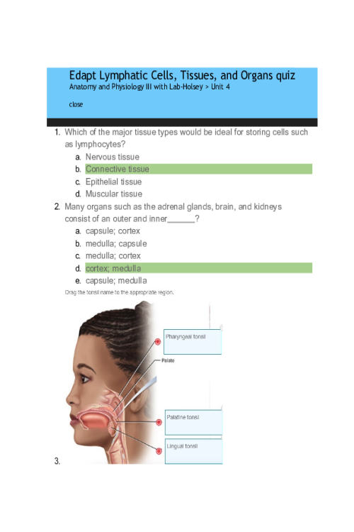
BIOS 255 Week 4 Concepts; The Lymphatic System (Edapt Lymphatic Cells, Tissues, Organs Quiz)
Contributor: Matthew Lillard
$10.00
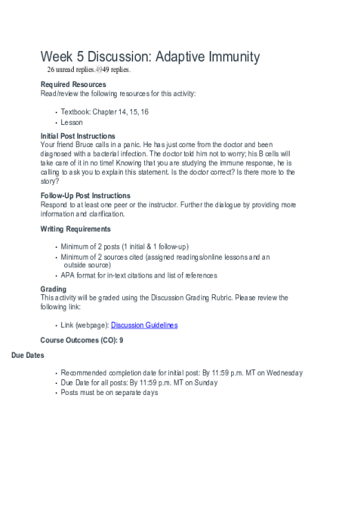
BIOS 255 Week 5 Discussion; The Lymphatic System and Immunity; Option A
Contributor: Matthew Lillard
$10.00
BIOS 255 Week 5 Virtual Lab; Introduction to Immunology Part I, Part II
Contributor: Matthew Lillard
$10.00
BIOS 255 Week 5 Assignment; Case Study I Hypersensitivity Reactions
Contributor: Matthew Lillard
$10.00
BIOS 255 Week 5 Concepts; The Immune System (Edapt Immune System Quiz)
Contributor: Matthew Lillard
$10.00
BIOS 255 Week 5 Concepts; The Immune System (Edapt Innate Defenses Quiz)
Contributor: Matthew Lillard
$10.00
BIOS 256 Week 2 Discussion; Peptic Ulcer Disease; A Case on the Digestive System.
Contributor: Daniel Sturridge
$10.00
BIOS 256 Week 2 OL Lab 2; Intestinal Glucose Transport; Study a mouse intestine model todiagnose an infant
Contributor: Daniel Sturridge
$20.00
BIOS 256 Week 3 Discussion; Glycogenesis, glycogenolysis, gluconeogenesis, and lipogenesis
Contributor: Daniel Sturridge
$10.00
BIOS 256 Week 3 OL Lab 3; Cellular Respiration; Measuring energy consumption during exercise
Contributor: Daniel Sturridge
$20.00
BIOS 256 Week 4 OL Lab 4; Renal Physiology; Find the mode of action of a diuretic drug
Contributor: Daniel Sturridge
$20.00
BIOS 256 Week 1 OL Lab 1; Carbohydrates The sugars that feed us
Contributor: Daniel Sturridge
$20.00
BIOS 256 Week 2 Discussion; Peptic Ulcer Disease; A Case on the Digestive System..
Contributor: Daniel Sturridge
$10.00
BIOS 256 Week 6 Discussion; male and female reproductive systems
Contributor: Daniel Sturridge
$10.00
BIOS 256 Week 7 OL Lab 7; Mendelian Inheritance From genes to traits
Contributor: Daniel Sturridge
$20.00
BIOS 256 Week 5 Discussion; Fluid balance, electrolyte balance, and acid-base balance
Contributor: Daniel Sturridge
$10.00
BIOS 256 Week 5 OL Lab 5; Fluid balance, acid base balance and water balance
Contributor: Daniel Sturridge
$20.00
